The PET scan makes use of a radioactive drug (tracer) to show both regular and abnormal metabolic activity. A PET scan can often become aware of the abnormal metabolism of the tracer in diseases earlier than the disease shows up on different imaging tests,
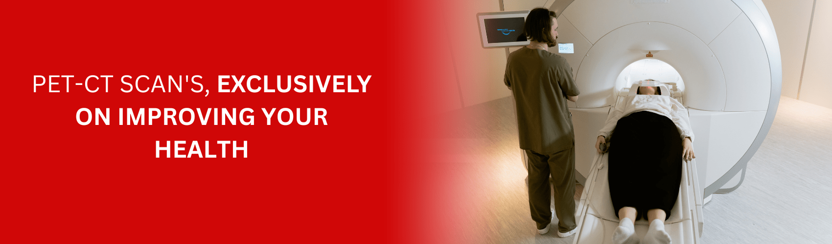
A PET Scan is a medical imaging procedure that combines a pet scan and a ct scan. A pet scan is a nuclear medicine imaging procedure that uses a camera to detect radiation from radioactive tracer injected into your body. A pet scan is a nuclear medicine imaging procedure that uses a camera to detect radiation from radioactive tracer injected into your body. The tracer is usually a radioactive sugar compound that contains a small amount of radioactive material.
The entire PET scan process takes about 60-90 minutes. It can also take up to 30 minutes for your body to absorb the injected radiotracer.
Do not consume or drink whatever, besides water, for six hours before the exam. You can drink water, as much water as you can would be helpful, till arrival. Routine medicines can be taken, until you have got been instructed otherwise.
A PET/CT scan can be more sensitive than other imaging checks and may find most cancers sooner than other tests do. Not all tumors absorb the radiotracer, however PET/CT is noticeably correct in differentiating from the benign and malignant tumors it reveals, mainly in some cancers such as lung and musculoskeletal tumors.
PET-CT Scan is a detailed investigation, our team of doctors will be reviewing and reporting your study within 24-48 working
hours.